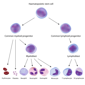
Blood-forming hematopoietic stem cells (top) give rise to all blood and immune cell types. In children with SCID, the steps leading to immune cells are broken.
In the world of fatal congenital immunodeficiency diseases, good news is always welcome, because most patients die before their first birthday if not treated. Babies with severe combined immunodeficiency disease, aka SCID or the “bubble boy disease,” now have more hope for survival thanks to two pieces of good news.
Credit: Samantha Morris, PhD, Boston Children's Hospital
If you’ve lost your way on the Boston subway, you need only consult a map to find the best route to your destination. Now stem cell engineers have a similar map to guide the making of cells and tissues for disease modeling, drug testing and regenerative medicine. It’s a computer algorithm known as CellNet.
As in this map on the cover of Cell, a cell has many possible destinations or “fates,” and can arrive at them through three main stem cell engineering methods:
• reprogramming (dialing a specialized cell, such as a skin cell, back to a stem-like state with full tissue-making potential)
• differentiation (pushing a stem cell to become a particular cell type, such as a blood cell)
• direct conversion (changing one kind of specialized cell to another kind)
Freely available on the Internet, CellNet provides clues to which methods of cellular engineering are most effective—and acts as a much-needed quality control tool. Full story »
At TEDx Longwood this spring, Leonard Zon, MD, founder and director of the Stem Cell Program at Boston Children’s Hospital, took the stage. In his enthusiastic yet humble style, he took the audience on a journey that included time-lapse video of zebrafish embryos developing, a riff by Jay Leno and a comparison of stem cell “engraftment” to a college kid coming home after finals: “You sleep for three days, and on day 4, you wake up and you’re in your own bed.” Three takeaways:
1) Stem cells made from our own skin cells can help find new therapeutics. With the right handling, they themselves can be therapeutics, producing healthy muscle, insulin-secreting cells, pretty much anything we need. (So far, this has just been done in mice.)
2) Zebrafish, especially when they’re see-through, can teach us how stem cells work and can be used for mass screening of potential drugs. The Zon Lab boasts 300,000 of these aquarium fish, and can mount robust “clinical trials” with 100 fish per group.
Zon, who co-founded the biopharm company Fate Therapeutics, will be part of a judging panel of clinicians and venture capitalists for the Innovation Tank at Boston Children’s Global Pediatric Innovation Summit + Awards (Oct. 30-31). Don’t miss it!
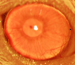
A restored, clear cornea grown from ABCB5-positive limbal stem cells. (Image courtesy of the researchers)
Severe burns, chemical injury and certain diseases can cause blindness by clouding the eyes’ corneas and killing off a precious population of stem cells that help maintain them. In the past, doctors have tried to regrow corneal tissue by transplanting cells from limbal tissue—found at the border between the cornea and the white of the eye. But they didn’t know whether the tissue contained enough of the active ingredient: limbal stem cells.
How cancer research led to a regenerative treatment for blindness.
Results have therefore been mixed. “Limbal stem cells are very rare, and successful transplants are dependent on these rare cells,” says Bruce Ksander, PhD, of the Massachusetts Eye and Ear/Schepens Eye Research Institute. “If you have a limbal stem cell deficiency and receive a transplant that does not contain stem cells, the cornea will become opaque again.”
Limbal stem cells have been sought for over a decade. That’s where a “tracer” molecule called ABCB5—first studied in the context of cancer—comes in. Full story »
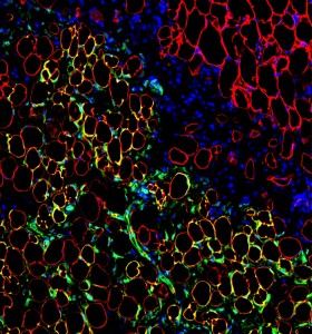
The fat cells shown in yellow are descended from transplanted human mesenchymal stem cells (green) inside of a mouse after co-transplantation. The red stain shows native mouse fat cells.(Courtesy Juan Melero-Martin)
Stem cell scientists had what first appeared to be an easy win for regenerative medicine when they discovered mesenchymal stem cells several decades ago. These cells, found in the bone marrow, can give rise to bone, fat and muscle tissue, and have been used in hundreds of clinical trials for tissue repair.
Uses range from tissue protection in heart attack and stroke to immune modification in multiple sclerosis and diabetes. Unfortunately, the results of these trials have been underwhelming. One challenge is that these stem cells don’t stick around in the body long enough to benefit the patient. Full story »
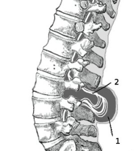
Prenatal cell therapy could avoid the need for invasive surgery to repair myelomeningocele.
The neural tube, which becomes the spinal cord and brain, is supposed to close during the first month of prenatal development. In children with spina bifida, it doesn’t close completely, leaving the nerves of the spinal cord exposed and subject to damage. The most common and serious form of spina bifida, myelomeningocele, sets a child up for lifelong disability, causing complications such as hydrocephalus, leg paralysis, and loss of bladder and bowel control.
New research from Boston Children’s Hospital, though still in animal models, suggests that standard amniocentesis, followed by one or more injections of cells into the womb, could be enough to at least partially repair spina bifida prenatally.
Currently, the standard procedure is to operate on infants soon after delivery. Full story »
The liver has been a model of tissue regeneration for decades, and it’s well known that a person’s liver cells can duplicate in response to injury. Even if three-quarters of the liver is surgically removed, duplication alone can return the organ to its normal functioning mass. It’s why people are able to donate part of their liver to someone in need—like this mother to her son who was born with biliary atresia.
But what about people with more chronic liver damage? Researchers led by Fernando Camargo, PhD, of the Harvard Stem Cell Institute and Boston Children’s Hospital’s Stem Cell Program, have new evidence in mice that it may be possible to repair such liver disease by forcing mature liver cells to turn back the clock and revert to a stem cell-like state, able to generate functional liver progenitor cells to replace damaged tissue. Full story »
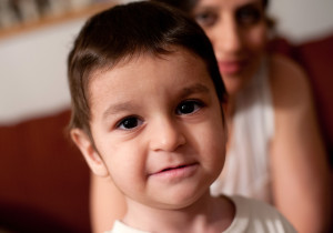
Emir Seyrek was the first patient with Wiskott-Aldrich syndrome to be treated in the U.S. in an international gene therapy trial.
Seeing that his mother, Kadriye, wasn’t looking, Emir Seyrek got an impish grin on his face, the kind only a two-year-old can have. He quietly dumped his bowl of dry cereal out on his bed and, with another quick look towards his mother, proceeded to pulverize the flakes to dust with his toy truck. The rest of the room burst out laughing while his mother scolded him. Despite the scolding, though, the impish grin remained.
It was the variability that intrigued pediatric cardiologist William Pu, MD, about his patient with heart failure. The boy suffered from a rare genetic mitochondrial disorder called Barth syndrome. While he ultimately needed a heart transplant, his heart function seemed to vary day-to-day, consistent with reports in the medical literature.
“Often patients present in infancy with severe heart failure, then in childhood it gets much better, and in the teen years, much worse,” says Pu, of the Cardiology Research Center at Boston Children’s Hospital. “This reversibility suggests that this is a disease we should really be able to fix.”
Though it needs much more testing, a potential fix may now be in sight for Barth syndrome, which has no specific treatment and also causes skeletal muscle weakness and low white-blood-cell counts. It’s taken the work of multiple labs collaborating across institutional lines. Full story »
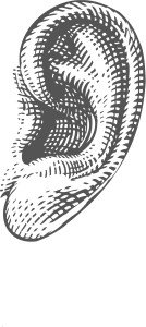
Hearing loss affects more than 300 million people worldwide, making it the most common sensory disorder. While there are no cures, recent efforts to develop biological treatments for hearing loss provide reason for cautious optimism. Three strategies—gene therapy, stem cells and drugs—have shown encouraging results in animal models, poising them for translation into potential therapies for humans.
Hearing loss can arise from many different causes, so it is unlikely that a single “magic bullet” will be developed to treat all forms of deafness. Rather, each individual cause may require a tailored and specific treatment strategy. Full story »