Just like Goldilocks wouldn’t eat porridge that was too hot or too cold, blood vessels won't grow properly in tissues that are too stiff or too loose. (Project Gutenberg/Wikimedia Commons)
In the tale Goldilocks and the Three Bears, Goldilocks tries all of the bears’ porridge, chairs and beds, finding that only the little bear’s things were just right. Everything else was a little off for her…too hot or too cold, too hard or too soft and so on.
Similarly, for everything to work as it should in the body, things need to be just right. Blood pressure shouldn’t be too high or too low; organs can’t be too big or too small, etc.
Donald Ingber, MD, PhD, and his lab in Boston Children’s Vascular Biology Program take this “just right” approach when thinking about how organs and tissues are structured. Recently, he and a member of his research staff, Akiko Mammoto, MD, PhD, discovered that by changing the stiffness of the surrounding tissues—not too loose and not too tight— they could keep blood vessels from leaking. Their finding could have real consequences for people with sepsis or other diseases featuring leaky vessels. Full story »
Clinical research is all about numbers. A new informatics network called SHRINE could help make it easier to get find out if the numbers of patients are there to answer complex questions. (victoriapeckham/Flickr)
Ed. note: This morning at 8:15 EDT, Isaac Kohane, MD, PhD, will tell the audience at TEDMED 2013 about his goal of using every clinical visit to advance medical science.
To preview his talk, we’ve updated a past Vector story about SHRINE, a system Kohane helped develop to allow scientists to use clinical data from multiple hospitals for research.
Clinical research really comes down to a numbers game. And those numbers can be the bane of the clinical researcher. If there aren’t enough patients in a study, its results could be statistically meaningless. But getting enough patients for a study, particularly for rare diseases, can be a daunting challenge.
The Shared Research Information Network (or SHRINE) could help solve this vexing problem. Developed through Harvard Catalyst by a team led by Isaac “Zak” Kohane, MD, PhD, director of Boston Children’s Hospital’s Informatics Program, SHRINE links the clinical databases of participating Harvard-affiliated hospitals—currently Boston Children’s Hospital, Beth Israel Deaconess Medical Center, Brigham and Women’s Hospital, Dana-Farber Cancer Institute and Massachusetts General Hospital—letting researchers at those hospitals see how many patients from those hospitals meet selected criteria.
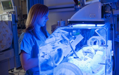
Babies with newborn jaundice need phototherapy. In the developed world that's easy; in the developing world, not so much. (Bruce R. Wahl/Beth Israel Deaconess Medical Center)
Family lore has it that when I was born, I had to spend a couple of extra days in the hospital for jaundice, the distinctive yellow tint to the skin that shows that a baby’s liver isn’t fully up and running yet. For me—and most of the newborns that develop jaundice every year in the developed world—the treatment was simple: spending some time lying under bright blue lights (aka phototherapy).
Note that I said “developed world.” The story in the developing world is quite different. Sometimes the nearest hospital with phototherapy equipment is hours’ or days’ travel away. Even though it’s simple, phototherapy is power intensive; no power, no treatment.
And untreated jaundice can have devastating consequences. The yellow pigment, called bilirubin, can accumulate in the brain and cause permanent brain damage or death.
The best solution for regions with few resources would have to be small and portable, run on batteries or other off-grid power sources, cost little, but still be safe and deliver the right wavelength and intensity of light. This is where Donna Brezinski, MD, wants to make a difference. And the Bili-Hut is her answer. Full story »
There’s no other way to say it: sepsis is a horrible disease. It typically starts with a runaway bacterial infection in the blood, followed by a runaway immune response that severely damages the body it’s trying to save. The results: shock, multiple organ failure and—in between 210,000 and 375,000 people in the United States alone every year—death.
Part of the problem is that the methods available for treating sepsis aren’t particularly good. Antibiotics can kill the bacteria, but that still leaves bacterial debris floating in the bloodstream, fueling the already over-excited inflammatory response.
Removing the bacteria altogether—as fast as possible—would be the better solution. At least that’s what Daniel Kohane, MD, PhD, thinks. His lab at Boston Children’s Hospital’s Division of Critical Care Medicine has developed a new approach that combines magnetic nanoparticles, a synthetic molecule (called bis-Zn-DPA) that binds to the bacteria, and magnetized microfluidic devices to pull bacteria from the blood quickly and efficiently. Full story »
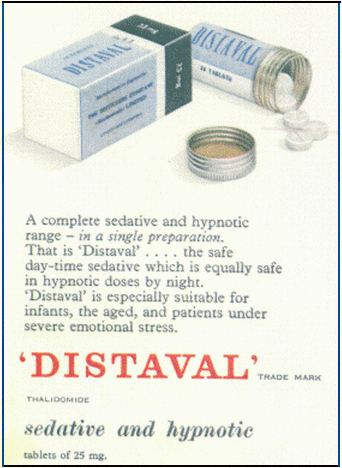
In times past, a pharmaceutical chemist’s main focus was to synthesize novel molecules to treat diseases. Today, an increasingly popular alternative is to re-engineer an existing drug—and continually improve it even after FDA approval. That’s how Robert D’Amato, MD, PhD, developed Pomalyst®, recently approved to treat multiple myeloma and the most potent analog to date of thalidomide.
Thalidomide has its own fascinating history. Originally developed by Chemie Grünenthal GmbH in the 1950s, it was the result of a search for an anti-anxiety drug to compete with Valium, and was approved for use in Europe as a sleep aid and depression treatment. Eventually, doctors found it useful for treating nausea, and started prescribing it off-label to pregnant women with morning sickness.
The results were disastrous. Thalidomide turned out to be a teratogen, causing severe birth defects. Full story »
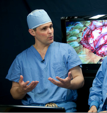
Ed Smith explains the moyamoya operation during a live webcast.
Lindsay Hoshaw contributed to this post.
It’s 7 a.m. and neurosurgeon
Ed Smith, MD, is downing a Diet Coke as he reviews the MRIs of today’s patients. He sprints up a stairwell to greet his first patient in the pre-operating wing.
Thirteen-year-old Maribel Ramos, about to have brain surgery at Boston Children’s Hospital, sits in her bed fidgeting. Smith reassures her about the operation, promises they’ll shave off as little hair as possible, and gets Maribel to crack a smile by telling her he moonlights as a hairdresser. Full story »
Recombinant DNA technology might turn bacteria into factories for producing siRNAs. (zoetnet/Flickr)
If you are a scientist and you want to turn off a gene, one option that’s been gaining traction is RNA interference (or RNAi). In this molecular process—first discovered in plants and only 12 years ago detected in mammals—bits of RNA called small interfering RNAs (siRNAs) cancel out a gene’s messenger RNA, effectively muffling that gene.
Labs can order custom-made, chemically synthesized siRNAs for just about any DNA sequence they want to silence. The tricky part is deciding what the right sequence is—especially when that gene is part of a virus, where genes can mutate pretty quickly.
However, a biotechnology approach to producing siRNAs could make it relatively easy for just about any lab that can master recombinant DNA technologies to make a number of siRNAs against multiple sequences within the same target gene: a potential bonus for companies seeking to make drugs that rely on RNAi. Full story »
A technology from a small research institute, originally developed as a safer way to make embryonic-like stem cells, just hooked a very large fish. As The New York Timesreported yesterday, pharma giant AstraZeneca is betting at least $240 million that this technology could be the source of a variety of new drugs—drugs that spur the body itself to make what it needs.
In 2010, the lab of Derrick Rossi at the Immune Disease Institute, which is now the Program in Cellular and Molecular Medicine at Boston Children’s Hospital, reported that they could reprogram ordinary cells into pluripotent stem cells by simply injecting them with messenger RNAs. The mRNAs reprogrammed the cells up to 100 percent more efficiently than other techniques, and did so without becoming part of the cell’s genome, greatly reducing concerns about cancer associated with other methods.
Key to the discovery were the chemical modifications made to the mRNAs so that cells wouldn’t “see” them as viruses and attack them. This video and this article describe the modified mRNA technique, also described in Cell Stem Cell:
Ed. note: Last week we wrote about Jurriaan Peters, MD’s brain network analysis in children with autism. In the second of our two part series on brain mapping, we talk about ways of mapping the brain’s physical wiring.
(AMagill/Flickr)
At the most basic level, the brain is a collection of wires, albeit a really complex one.
But how during development do nerve fibers thread their way through the growing brain and make the right connections?
“We know very little about what’s happening in the developing brain in three dimensions,” says Emi Takahashi, PhD, a researcher in the Fetal-Neonatal Neuroimaging & Developmental Science Center (FNNDSC) at Boston Children’s Hospital. “With histology techniques, we can achieve a two-dimensional view over small areas, but it’s hard to know which fiber bundles are growing in which ways during different stages of development in the whole brain.”
But new MRI-based technologies are quickly closing that knowledge gap, giving us our first high-resolution peek into how the developing brain wires itself up.
Using something called high angular resolution diffusion imaging (HARDI) MRI, Takahashi and her colleagues (including neuroradiologist and FNNDSC director P. Ellen Grant, MD) can trace the three-dimensional pathways within the growing brain via stunning images like these:
Sharing via social media is a great opportunity for collecting better public health data and encouraging healthy behavior changes. (bengrey/Flickr)
We humans are sharing creatures. We talk about ourselves, what we think, what we know. If we weren’t like this, cocktail parties would be really boring, and Facebook and Twitter wouldn’t exist.
Nor would health care. At the most basic level, health care relies on give-and-take between patients and doctors—patients sharing their symptoms and concerns with doctors, and doctors sharing their knowledge with patients.
The same holds true for public health. Prevention and control efforts require lots of patients and doctors to share information so that public health agencies know where to target their resources.
But the give-and-take in public health is often slow and cannot always detect conditions or complications at rates that reflect reality. And usually it’s one-way—from the patient or public to surveyors. Full story »