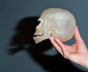
 A picture may be worth a thousand words, but there’s something about holding an object in your hands that’s worth so much more. I realized this when John Meara, MD, DMD, handed me the skull of one of his patients.
I turned it over in my hands while Meara, Boston Children’s Hospital’s plastic surgeon-in-chief, pointed out features like the cranium’s asymmetric shape and the face’s malformed left orbit.
Mind you, it wasn’t actually Meara’s patient’s skull in my hands. In reality, I was holding a high-resolution, plastic 3D model printed from the patient’s CT scans.
The printer that made that model—and several other models I saw in the last month—is the centerpiece of a new in-house 3D printing service being built by Peter Weinstock, MD, PhD, and Boston Children’s Simulator Program.
3D printing technology has exploded in the last few years, to the point where anyone can buy a 3D printer like the MakerBot for a couple of thousand dollars or order 3D printed products from services like Shapeways. Adobe even recently added 3D printing support to Photoshop.
And 3D printing is already making a mark on medicine. Just look at stories like that of a baby whose windpipe was replaced with a 3D-printed replica, or a U.K. patient who now has a 3D-printed pelvis.
Instead of printing implants, Weinstock and his team are putting a child’s internal anatomy in surgeons’ hands before going near an operating room.
A new level of simulation
Weinstock, who is also a critical care physician, views the service as a further step in the evolution of medical simulation and a way of linking preparedness inextricably with patient care. “It started with using a mannequin in an office,” he says. “We brought it into the hospital and started using higher fidelity mannequins that could enhance fellowship training. Step 3 was bringing simulation to the point of care, so that teams could train and practice together.”
“With 3D printing, we’re taking a step that allows experienced doctors to simulate the specific anatomy of their patients and allows the best of the best become even better,” he continues. “We’ve created a continuum of simulation that provides different levels of practice depending on the expertise and needs of the trainee.”
Not your average 3D printer
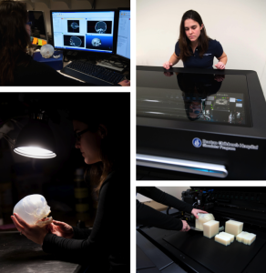
The making a model (clockwise from top left): Importing a patient's images into a 3D design program for printing; checking the print job; taking the model, embedded in a removable support material, out of the printer; examining the cleaned and finished product.
Second, the printer—provided by the hospital’s Department of Anesthesia, Perioperative and Pain Medicine—has extremely high resolution: down to 16 microns (the size of a droplet in a cloud). This allows models to capture fine details, which can be critical for something as small as a baby’s brain or skull.
Third, the models can be built from multiple plastics, mimicking the physical characteristics of different tissues like bone, skin and blood vessels all in a single model.
Lastly, because the printer uses individual patients’ CT or MRI scans as source data, the models accurately capture the unique anatomy of each individual child.
“We see it as the realization of ultra-high fidelity simulation,” says Weinstock. “Imagine being able to print an organ and surrounding anatomy, take it into the simulation suite or the operating room and practice on it. Then take that same model you’ve operated on and use it as a guide for treating that child.”
“This could be a game changer in surgical preparedness,” he adds.
Your child’s model is ready
Neurosurgeon Joseph Madsen, MD, expresses a similar sentiment as he reaches into his desk and pulls out a printed model of a brain based on one of his epilepsy patients. The model shows her brain’s exact contours, even the placement of her “grid”: the network of electrodes Madsen’s team used to find the focal point of her seizure activity.
“It can be hard to conceptualize and explain an operation just from imaging,” says Madsen, who with engineer Tomer Anor, PhD, is using 3D printing to develop and train neurosurgical robots. “In a case like this, there’s no good way to practice before surgery.”
Except to practice with a model, as Madsen has done for another epilepsy patient who needed to undergo a hemispherectomy (removal or disconnection of one hemisphere of the brain). “Doing a dry run of the case ahead of time using a model from this child’s imaging has certainly helped us,” he says.
Neurosurgeon Joseph Madsen, MD, with a model brain printed from images of one of his patients. Notice the different plastics for different brain structures.
Darren Orbach, MD, PhD, and his colleague Edward Smith, MD, are already looking at turning simulation into a regular clinical offering, making 3D-printed models a standard service for every patient who comes to their recently launched Cerebrovascular Surgery and Interventions Center.
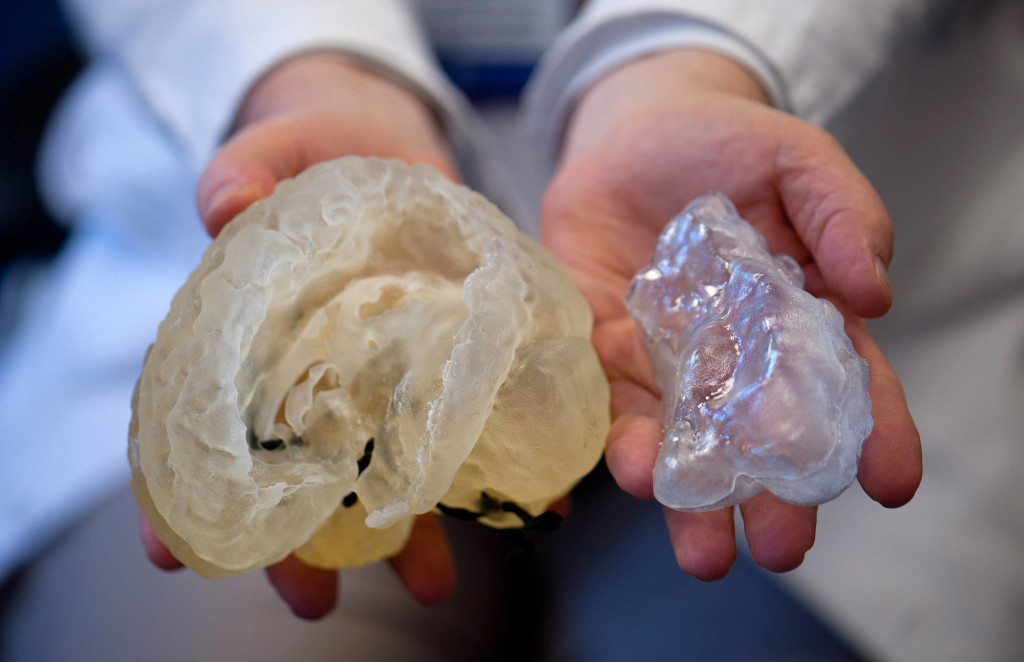
“It’s very different to hold a model in your hands when you’ve been used to trying to manipulate images on a screen for years,” says Orbach, a neurointerventional radiologist, showing me 3D-printed blood vessels from the brain of a child with a vein of Galen malformation. He thinks that models with hollow veins and arteries—something he’s discussing with Weinstock—could have great potential for practicing vascular procedures. “They would let me figure out directly what approaches and catheter sizes would work and what wouldn’t before going into the catheterization suite.”
Smith, the hospital’s director of cerebrovascular neurosurgery, envisions practicing surgery on a patient’s model and using a scan of that model as a real-time guide in the operating room.
Interventional radiologist Darren Orbach, MD, PhD, holds a model from a patient with a rare vascular anomaly called a vein of Galen.
Tool for the next generation
Weinstock’s models also may be important tools for training future specialists.
Meara points to his office shelves. They hold models of skulls purchased from a commercial vendor, each an example of a different craniofacial anomaly. While those models have some training value for new fellows, they don’t capture the breadth of variation he sees in his practice. “Each of those models represents an ‘average’ for each anomaly. But every child’s case is different.”
Nor can the bought models be operated on. Made with an older technology, they don’t have the material properties of bone and tissue and can’t be cut or manipulated like Weinstock’s models.
“It’s hard for a trainee to visualize exactly what a severe anomaly looks like,” Meara says. “To give them the opportunity to hold and feel the anatomy lets them train their proprioceptive senses and learn more than they could from imaging.”
Orbach agrees, “If we could make models tailored specific cases and specific trainees, it could be revolutionary.”
“I have more fellows to train than I have patients who need hemispherectomies,” adds Madsen. “But there’s no reason we couldn’t make a model, operate on it, scan it to record what we did and use it as the basis of a training module.”
Opportunities for creativity
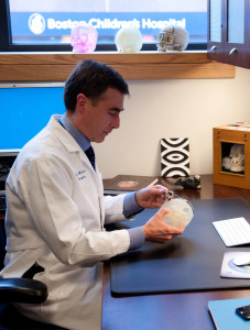
Plastic surgeon-in-chief John Meara, MD, DMD, measures features of a model of a patient's skull.
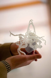
“We had already completed this child’s surgery and asked for the models, so that the next time we saw this kind of anomaly we’d be more prepared,” Afshar says. The spiral cuts were one of several surgical solutions Afshar and his colleagues came up with. “It’s an approach we wouldn’t have thought to try based on just imaging alone.”
“It’s the rare kind of case that might come in once or twice a year,” Meara adds. “With the models, we can try 10 different approaches on the same case without risking harm to a patient.”
“It’s good to see how 3D printing inspires people and gets them to come up with new ideas about how to approach patient care,” says Madsen. “We’re used to thinking about anatomy in one way, and then someone like Peter comes along and says, ‘Let’s print that.’ It brings a new perspective that can help solve old problems.”


