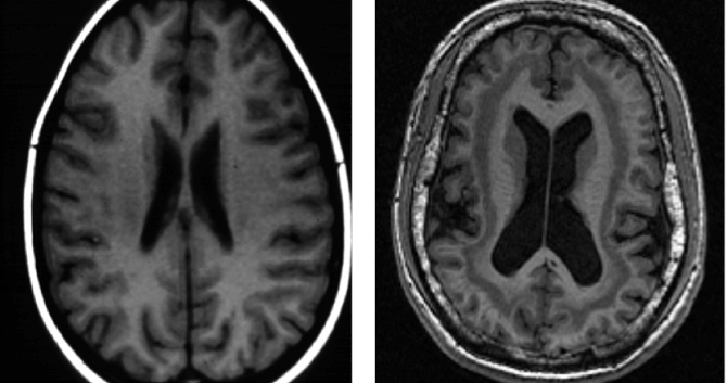
 (Above: In double cortex syndrome, causing epilepsy and mental retardation, an extra cortex forms just beneath the cerebral cortex [right]. The causative DCX mutation interferes with migration of neurons during the cortex’s early development. Courtesy Walsh Lab)
The journal Neuron, celebrating its 25th anniversary, recently picked one influential neuroscience paper from each year of the publication. In this two-part series, we feature the two Boston Children’s Hospital’s scientists who made the cut. The Q&A below is adapted with kind permission from Cell Press. (See part 1)
Key to well-tuned brain function is the migration of neurons to precise locations as the brain develops. The long journey begins deep inside the brain and ends in the outer cerebral cortex—where our highest cognitive functions lie. Christopher Walsh, MD, PhD, has shown that several genetic mutations causing neurodevelopmental disorders disrupt this neuronal migration, landing neurons in the wrong places. Each gene governs a specific sub-task: one kicks off the migration process; others stop migration when neurons have arrived in the right location. Full story »
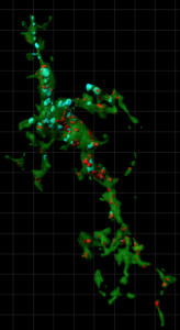
Caught in the act: This microglial cell receives visual input from the eyes. The blue and red dots (inputs from the same-side and opposite-side eye, respectively) are synapses the cell has engulfed—the first step in eliminating synapses the brain no longer needs.
The journal Neuron,
celebrating its 25th anniversary, recently picked one influential neuroscience paper from each year of the publication. In this two-part series, we feature the two Boston Children’s Hospital’s scientists who made the cut. The Q&A below is adapted with kind permission from Cell Press.
In 2012, Beth Stevens, PhD, and colleagues provided a new understanding of how glial cells shape healthy brain development. Glia were once thought to be merely nerve “glue” (the meaning of “glia” from the Greek), serving only to protect and support neurons. “In the field of neuroscience, glia have often been ignored,” Stevens told Vector last year.
No longer. Stevens’s 2012 paper documented that microglia—glial cells best known for their immune function—are no passive bystanders. They get rid of excess connections, or synapses, in the developing brain the same way they’d dispatch an invading pathogen—by eating them. Full story »
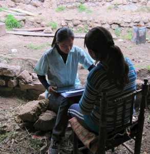
A fieldworker in Peru administers cognitive tests to a 12-year-old girl. While socioeconomic circumstances can vary dramatically, how families perceive and adapt to them may be more critical in brain development.
Studies going back to the 1950s have linked objective socioeconomic factors—like parental income or education—to child health and achievement. Recent studies have extended this research, indicating that parental socioeconomic status (SES) also
affects physiologic brain function in children. A
new study, while small, is the first to suggest another potent factor: the mother’s
self-perceived social status
Margaret Sheridan, PhD, at Boston Children’s Hospital’s Labs of Cognitive Neuroscience, and colleagues studied 38 children, ages 8 to 12 years. Each child gave a saliva sample to measure levels of the stress hormone cortisol, and 19 also underwent functional MRI of the brain focusing on the hippocampus, a structure responsible for long-term memory formation (required for learning) and for reducing stress responses.
Their mothers, meanwhile, were shown a picture of a ladder and were asked to rank their social status on a scale of 1 to 10 as compared with others in the United States. Full story »

Illustration: David Roberson
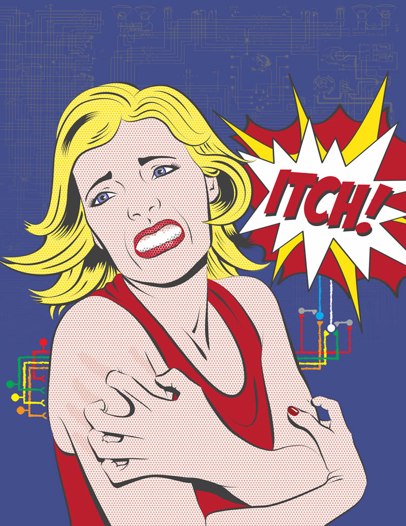
This post originally appeared in longer form on Harvard Medical School’s news site. Try not to scratch when you read it.
There’s itch, and then there’s itch.
New research has revealed distinct sets of itch-generating neurons that explain why current itch therapies often fail. It also suggests new ways to selectively silence itch.
“We think this [research] has therapeutic implications,” says Clifford Woolf, PhD, director of the F.M. Kirby Neurobiology Center at Boston Children’s Hospital and professor of neurology at Harvard Medical School (HMS).
While itch is more aggravating than life-threatening, Woolf and HMS graduate student David Roberson hope their work might one day ease the torment itch can cause, particularly in children.
“If you go into the pediatric immunology wards, you see little kids with their hands in mittens or sometimes tied down because they scratch themselves to a point where they damage themselves,” says Woolf. Full story »

Crossing the river has had benefits that go back decades (Roger Wollstadt/Flickr, 1975)
It’s inspiring to see what happens when a hospital dedicated to providing the best treatments for children partners with a world-class technology and engineering institution. Children’s Hospital Boston and MIT have embarked upon an exciting program of collaboration and cross-fertilization in research, teaching and mentoring. The goal is to connect outstanding disease-oriented research with cutting-edge innovation and technologies, taking our ability to care for children to a new level while training the next generation of clinicians and scientists.
The historical ties between Children’s and MIT run deep. Individual scientists and clinicians have teamed up to design new medical devices; to identify gene mutations that underlie cancer and disorders of development; to create new approaches to drug delivery using slow-release polymers to extend medication efficacy; Full story »

(luisar/Flickr)
What if blind eyes could see? What does that mean?
That’s the question neuroscientist Pawan Sinha and his team at MIT has begun to answer in a uniquely humanitarian and scientific endeavor.
Project Prakash (named for the Sanskrit word for “light”) intended, at first, to cure blind children in India. It’s a noble effort, given that India has the world’s highest population of blind people, less than half of whom survive to their third birthday and less than one percent of whom are employable.
Sinha’s team screened 20,000 blind Indian children and treated 700 of them for correctable problems such as cataracts. As Sinha recounted at last month’s One Mind for Research forum, these 700 children now can see.
Sort of. Full story »

Illustration of the pain pathway in René Descartes' Traite de l'homme (Treatise of Man), 1664. The long fiber running from the foot to the cavity in the head is pulled by the heat and releases a fluid that makes the muscles contract.
Christopher Walsh, MD, PhD, is chief of Genetics and a Howard Hughes Medical Institute Investigator at Children’s Hospital Boston, where his research focuses on genes that regulate the development and function of the human cerebral cortex. These genes are vital to normal development of the cortex, and many appear to have been altered evolutionarily to allow the unique aspects of the brain that underlie human cognitive abilities. Mutations in these genes are known to cause autism and epilepsy, as well as intellectual disabilities and other learning disorders.
In the second century, the Greek physician Galen proposed that the fluid in the brain provided energy for the entire body, theorizing that an external spirit (pneuma) from the lungs was transported to the heart, where, combined with blood, it would give rise to the vital spirit. Carried by the blood, the vital spirit was then thought to be transformed into an animal spirit before entering the cavities of the brain, then traveling through the nerves, “as sunshine passes through the air or water,” to energize the entire physical being.
In this view, the brain itself was a mere holder of the fluid. Our neuroanatomical term “thalamus,” referring to part of the brain stem, comes from the Greek word for “chamber,” implying that the brain was mainly important as a holder for CSF. The philosopher Descartes (1596-1650) thought that the brain was a pump that moved the fluid around to do the brain’s work, such as making a muscle contract. Seventeenth century Swedish scientist Emanuel Swedenborg referred to the CSF as a “spirituous lymph” and a “highly gifted juice.”
Full story »

Eye muscles, the nerves that control them, and where things go wrong (click to enlarge)
Elizabeth Engle used to wait in peoples’ driveways until midnight, hoping to enroll them in her genetic studies of eye-movement disorders. She landed there by chance: during her neurology residency, she saw a little boy whose eyes were frozen in a downward gaze. Wanting to find a solution to a disorder that others had written off, she talked her way into the muscular dystrophy genetics lab of Alan Beggs and Lou Kunkel at Children’s.
Why muscular dystrophy? That tragic muscle-weakening disease somehow spares the eye muscles. Engle thought if Beggs and Kunkel took her on, she could answer two questions at once – what was protecting the eye muscles in muscular dystrophy, and what had caused the little boy’s fixed gaze and droopy eyelids. Plus, she needed laboratory training to study the samples she’d started gathering. “I didn’t have a PhD and was never officially trained in the lab,” she once said. “I didn’t even know how to make chemical solutions.” Full story »