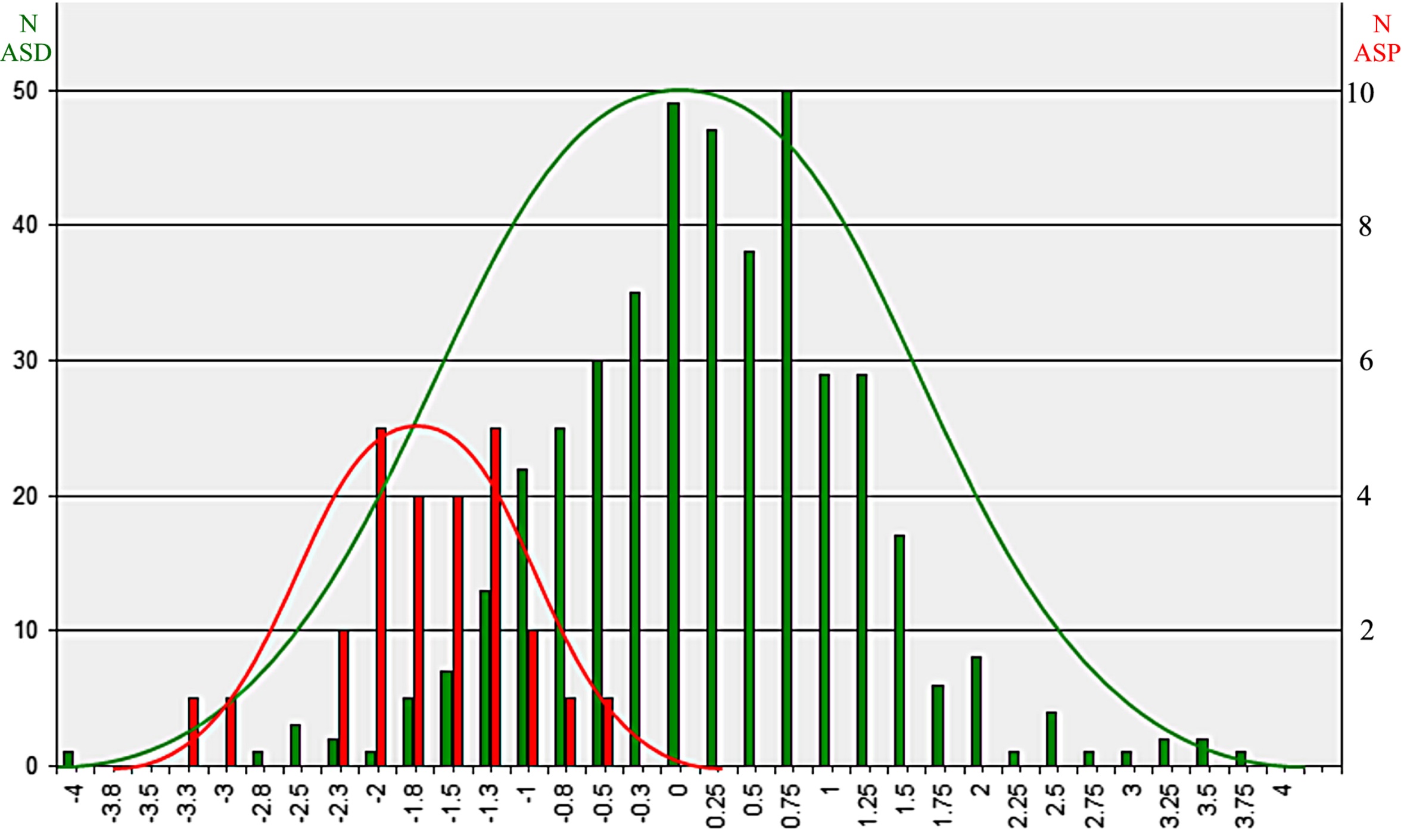
Asperger’s syndrome vs. autism spectrum disorders:
This histogram separates children with Asperger’s (in red) from those with autism spectrum disorders (green) based on EEG coherence variables. Although there is overlap with high-functioning autism, the Asperger’s children clearly form a distinct group. (Courtesy BMC Medicine)
Is it Asperger’s syndrome or is it autism? Since there are no objective diagnostic measures, the diagnosis is often rather squishy, based on how individual clinicians interpret a child’s behavior. According to the Diagnostic and Statistical Manual, fourth edition (DSM-IV), early problems with language development are an indicator of autism; if there are behavioral symptoms but no early language problems, the child has Asperger’s. However, if the diagnosis is made late, parents’ recall of early language development may be fuzzy.
Under the new DSM-V, published in May, Asperger’s is included under the general “autism spectrum disorders (ASD)” umbrella. This has raised concerns among families who feel their children with Asperger’s have unique needs that won’t be met in classroom programs designed for autism.
Frank Duffy, MD, a neurologist at Boston Children’s Hospital, believes it’s possible to objectively differentiate Asperger’s from ASDs using a new wrinkle on an old technology. Originally trained as an engineer, Duffy is expert at interpreting electroencephalography (EEG) signals—the wiggly lines that represent electrical activity in the brain. Full story »
Seizures seem to strengthen and "lock in" synapses too soon, leaving no room for development. (Image: Ice synapses, Joe Flintham/Flickr)
It’s well known that babies who have seizures soon after birth have roughly a 50-50 chance of developing long-term intellectual and memory deficits and cognitive disorders like autism. But until now, it wasn’t understood why these deficits occur, much less how to prevent them from happening.
In the December 14 Journal of Neuroscience, researchers at Children’s Hospital Boston, led by neurologist-neuroscientist Frances Jensen, detail in a rat model how early-life seizures affect brain development at the cellular and molecular level. But more to the point, they show that it might be possible to ward off these effects with drug treatment soon after the seizure – using a drug called NBQX or similar drugs that are already approved by the FDA.
Jenson was particularly interested in what seizures do to synapses, the connections between neurons that are rapidly developing in the infant brain.
Children’s neurologist-neuroscientist Mustafa Sahin, Simon Warfield, director of the hospital’s Computational Radiology Laboratory, and Jurriaan Peters compared brain organization in 29 healthy subjects with that in 40 patients with tuberous sclerosis, a rare genetic syndrome often associated with cognitive and behavioral deficits, including ASDs about 50 percent of the time. “Patients with tuberous sclerosis can be diagnosed at birth or potentially before birth, because of cardiac tumors that are visible on ultrasound, giving us the opportunity to understand the circuitry of the brain at an early age,” explains Sahin.
The panels above (click to enlarge) are advanced MRI images Full story »
A sequence of motion frames of a normally kicking baby's legs (shown in blue and green), illustrating changing joint angles at the hip and knee.
Countless scientific epiphanies never leave the bench – unless there’s the kind of serendipitous encounter that set Children’s Hospital Boston psychologist Gene Goldfield on a path he never expected to follow.
One in eight babies are born prematurely, putting them at greater risk for cerebral palsy, an inability to fully control their muscles. Goldfield saw these children being wheeled around the hospital, and was convinced that they did not have to be wheelchair-bound.
During early infancy, he knew, the developing brain naturally undergoes a rewiring of its circuits, including those that control the muscles. Could some type of early intervention encourage more typical motor development by replacing damaged circuits with more functional connections?
At Children’s Innovators’ Forum last week, Goldfield discussed his envisioned solution: the use of programmable robots Full story »
The right inferior frontal gyrus, part of the prefrontal cortex, lights up on fMRI when children play a game requiring them to resist a natural impulse. This brain area is naturally in flux between ages 5 and 7, Sheridan has found.
Last month, the American Academy of Pediatrics released new guidelines on attention-deficit hyperactivity disorder (ADHD), lowering the minimum age at which physicians should consider drug treatment from 6 years to 4 years.
But here’s the problem. “Current behavioral criteria for ADHD are most effective only after age 8 or 9,” says Margaret Sheridan of the Laboratories of Cognitive Neuroscience at Children’s Hospital Boston. “If you use them at age 3 to 6, then you’re wrong about half the time, and the child will stop meeting the criteria by age 8.”
Little kids, especially boys, are naturally distractible, impulsive and fidgety. Some mature more slowly; some are just the youngest in their class. Many will grow out of their wild but largely age-appropriate behaviors.
But letting true ADHD fester, explaining symptoms away as “kids just being kids,” deprives children of the benefits of behavioral or pharmacologic treatment at a time when their young brains are highly responsive. Full story »
Mitochondria, as you may know, are the engines that power cells. They’re always in motion, supplying energy wherever it’s needed. In brain cells, mitochondria especially have to hoof it around, traveling out into the axons and dendrites to fuel the energy-intensive task of communicating with other cells.
But in at least one form of Parkinson’s disease, that movement becomes a problem: the genetic mutations causing the disease leave neurons unable to make the fidgety organelles hold still. Without this ability, the dopamine-producing neurons in the brain’s substantia nigra can’t safely dispose of mitochondria when they go bad, and the neurons die or become impaired.
“When damaged, mitochondria produce reactive oxygen species that are highly destructive, and can fuse with healthy mitochondria and contaminate them, too,” explains Tom Schwarz, of the F.M. Kirby Neurobiology Center at Children’s Hospital Boston, senior investigator on a study published in Cell today. “It’s the equivalent of an environmental disaster in the cell.” Full story »
Evacuation of a soldier injured by a roadside bomb, June 17, 2011, Kandahar province of Afghanistan (DVIDSHUB/Flickr)
From the time he was 11, Robert Tasker knew he wanted to be a doctor. The son of a serviceman, he was drawn to battlefield surgery, evacuations and managing traumatic injuries. Instead, he ended up on a different kind of battlefield, where what’s at stake are the highly vulnerable, still developing brains of infants and children – and where it’s critical to be mobile and show up on time.
Tasker directs the Pediatric NeuroCritical Care program at Children’s Hospital Boston, the first of its kind in the world. His goal is to protect brain function not only in children suffering direct head injury, but children undergoing major surgery, children with stroke, children hospitalized for critical illness, children on ventilators, children with nervous-system infections like meningitis and more.
Born in Hong Kong and raised throughout the globe, Full story »
Patients with severe epilepsy can have seizures every day – sometimes waking up on the floor, not knowing what happened. For about 1 in 3 epilepsy patients, drugs are of no help. An implanted vagus nerve stimulator can sometimes control seizures, but often not. Surgically removing the excitable brain tissue can be curative, but often, too many areas of the brain are involved to effectively remove the entire seizure focus. Or the area causing the seizures is too close to a vital brain area – say, a memory or motor area – making surgery too risky.
Alex Rotenberg, a neurologist in Children’s Hospital Boston’s epilepsy program, has been having success with an experimental technique for this kind of disabling, treatment-resistant epilepsy. Known as repetitive transcranial magnetic stimulation (rTMS), it has helped a small number of patients with no other good options for controlling their seizures, such as Kate:
Benjamin Warf, MD, director of Neonatal and Congenital Anomalies Neurosurgery at Children’s Hospital Boston, developed a new treatment for infant hydrocephalus, or “water on the brain,” while a medical missionary in Africa, where hydrocephalus is common and usually untreated. His innovation, which has saved the lives of thousands of children, is minimally invasive, relatively inexpensive and has been taught to other surgeons in developing countries. The post below is adapted from Warf’s testimony last week before the House Subcommittee on Africa, Global Health and Human Rights (viewable on C-SPAN; jump to 17:54). John Mugamba, MD, whom Warf trained and who is currently medical director at CURE Children’s Hospital of Uganda, gave testimony in video form.
In 2000, my family and I moved to Uganda as medical missionaries to help start a specialty hospital for pediatric neurosurgery, the CURE Children’s Hospital of Uganda. At the time, there were no pediatric neurosurgical hospitals and few trained neurosurgeons in all of Africa. Full story »
Mark Bear’s research interests have taken him from studying vision in kittens to learning and memory in mouse models, and more recently, to the study of Fragile X syndrome, one of the leading genetic causes of autism and intellectual disability in humans. Along the way, he has made several ground-breaking contributions to neuroscience – one of which he described as one of MIT’s presenters at this week’s inaugural CHB-MIT Research Enterprise Symposium, which kicked off an exciting new scientific collaboration between MIT and Children’s.
I have followed Mark Bear’s work since I was an undergraduate at Brown University, where he used to teach the Introduction to Neuroscience course. That’s where I first learned about the seminal experiments in kittens (see this PDF), showing that covering one eye at birth rewires their brains not to “see” out of that eye, work that Bear was continuing to refine. Our paths crossed again more recently due to our common interest in studying autism. Full story »