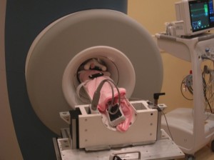
A 4.2-lb baby girl in the new 1.5 Tesla MRI magnet, designed for use in the NICU. (Images courtesy of Cincinnati Children’s Hospital Medical Center)
Experience suggests that magnetic resonance imaging (MRI) and advanced MR techniques such as spectroscopy and diffusion imaging offer substantial benefits when diagnosing problems in premature babies. However, today’s MR systems poses significant logistical barriers to imaging these infants. We have been working to change that.
MRI provides an unparalleled ability to visualize anatomy without the hazards of ionizing radiation. Yet premature and sick babies in neonatal intensive care units (NICUs) are usually too delicate to leave the unit. The few babies who receive MRI today must be accompanied by NICU staff during transport to and from the Radiology Department. This process is often a multi-hour ordeal and reduces the staff available to care for other babies in the NICU. Moreover, infants must be imaged in an adult-sized MRI scanner that has imaging coils designed for adult anatomy and a long magnet tunnel that prevents direct observation of infants.
Bringing MR down to size
To overcome these challenges, we created a new type of MR scanner—one small enough to be placed directly in the NICU. To do this, we converted a small-bore magnet designed for imaging adult knees into one suitable for imaging an infant’s whole body. Our 1.5 Tesla MR scanner employs the same strong magnetic field used in most MR systems but has a footprint of only a few square feet.
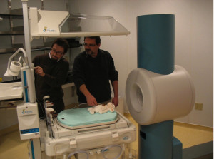
Engineers Wolfgang Loew (left) and Randy Giaquinto testing system interactions between the new magnet and a standard incubator.
Our new NICU MR system also incorporates new patient handling systems that address the special needs of infants, including those of premature babies and sick newborns. Its small size offers advantages over large-bore systems, such as:
- a substantial reduction in noise
- improved view of the baby while in the scanner
- the capability to bring support equipment such as ventilators and pumps closer to the baby during scanning
Launch experience
We tested our new NICU MR scanner in a pilot study of 15 healthy babies at Cincinnati Children’s in early 2012. The images matched or exceeded those possible with a conventional MRI system, and our average transport time (from bed to scanner and back) was less than 30 minutes. With this success, we began using the system to regularly diagnose conditions commonly found in newborns, and we have scanned more than 200 babies to date.
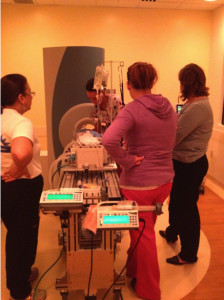
Staff members preparing a premature infant for scanning in Cincinnati Children’s NICU. Note that the baby is connected to a ventilator, two infusion pumps and an MR-compatible vital signs monitoring system.
Learning experience
We’ve learned several lessons with the new system. First, it has greatly reduced the logistical burden of scanning infants, making MR more accessible to the babies who need it the most. The scanner’s image quality and performance matches—and for some studies exceeds—that of larger scanners.
Secondly, as our experience with neonatal MRI increases, it is becoming apparent that the imaging methods traditionally employed in the NICU—x-ray and ultrasound—are lacking. Ultrasound’s use is limited to a few acoustic windows in the body, while x-ray imaging employs ionizing radiation. Neither imaging modality is well suited for visualizing soft tissue. Features that are difficult to evaluate with x-ray and ultrasound, such as bowel wall inflammation and abscesses, particularly in the presence of a bowel gas, are easily seen with MR.
MR’s superior power at detecting small hemorrhages in the brain also has called into question the utility of ultrasound for revealing brain injury in small babies. With the new MR system, we have diagnosed brain lesions that were inconspicuous on ultrasound. Because we can be more diagnostically certain with MR, we can react more quickly in acute cases and capture objective data for patient management both during infants’ stay and after discharge.
The future of neonatal MRI
While most scans we have performed to date have been on the brain, we are ramping up research programs in lung and abdominal imaging. For the first time, we can envision bringing the full power of diagnostic MRI to bear on common problems in neonatal medicine, such as respiratory distress syndrome, malformations of the heart and great vessels and necrotizing enterocolitis. The Clinical Innovation award from the National Pediatric Innovation Summit will help us push the scanner’s technical capabilities, including the creation of even quieter strategies for scanning sleeping babies.
Although ultrasound and x-ray imaging systems are available in most NICUs today, MRI has so much more to offer. We hope that in a few years, NICU MR imaging systems like the one in Cincinnati, designed to meet the needs of premature babies, will be available to NICU patients around the world. We finally have an MRI system that can bring the full power of this remarkable technology to our most vulnerable patients.