 It’s music to the ear of any cancer patient: “Your scans are clear.” It means you’ve won, and the treatments you’ve endured have driven cancer from your body.
But once you hear those four magic words, are you also free of the need for future scans? There’s an argument to be made that continued scanning or surveillance imaging is a good thing. After all, if you have a relapse, you want to detect it as early as possible.
But that argument may not hold up in the face of data. Continued imaging can be expensive, can expose survivors to additional radiation, can have false positive results leading to additional worry and unnecessary medical care, and may not be any better at detecting tumor relapses than a physical exam or simply a survivor’s feeling that something is “wrong.”
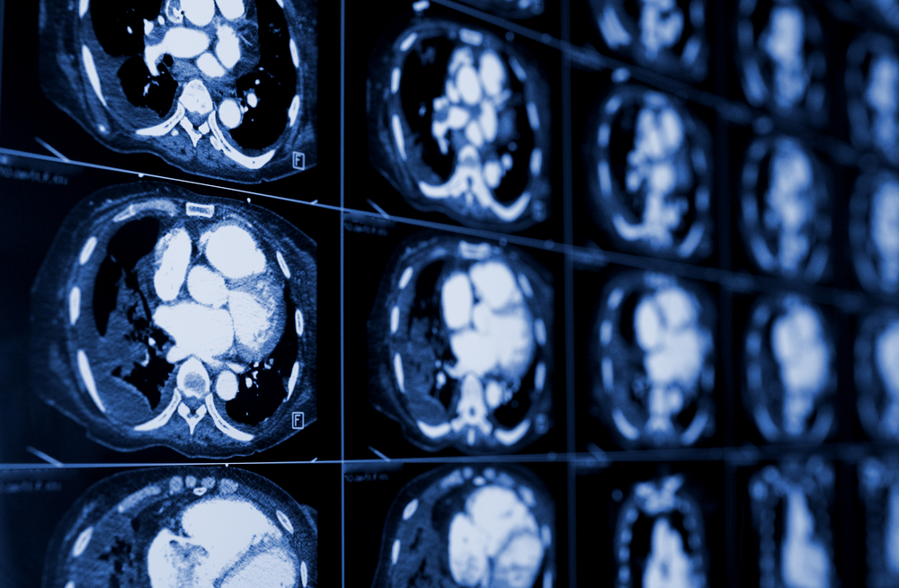
Stephan Voss, MD, PhD, the director of Nuclear Medicine and Molecular Imaging and chief of Oncologic Imaging at Boston Children’s Hospital, decided to crunch the numbers, using Hodgkin lymphoma (HL) as a model for testing whether post-treatment surveillance with computed tomography (CT) scans makes clinical sense. His conclusion: not really.
“The conventional wisdom is that early detection of relapse means that we spare the patient side effects and poorer outcomes,” Voss says, referring to the belief that HL survivors should have a follow-up CT scan every year for up to five years after treatment. “But with Hodgkin disease, that’s not the case.”
“Little influence on overall patient outcome”
But as Voss has pointed out in papers in the Journal of Clinical Oncology and Current Hematologic Malignancy Reports, few HL survivors relapse, and the majority of those who do relapse do so within the first year after treatment. “In the case of relapses that occur outside that one-year period, imaging has no impact on survival,” he says. “Patients tend to do quite well on second-line therapy.”
Or, as he writes in the Current Hematologic Malignancy Reports paper:
The data that has emerged over the past five years has indicated that routine disease surveillance either with CT or with PET/CT has little influence on overall patient outcome.
There’s also the issue of cost. Voss highlights a study by Dana-Farber Cancer Institute and Harvard University, in which doctors had to conduct $110,000 in CT scans to detect a single relapse in a population of HL survivors.
Voss also raises concerns about the false-positive rate of CT for HL detection—as high as 81 percent in one of the studies he cites. “If you use a test like CT that’s good at picking things up, you are almost certain to find something, whether or not it’s relevant. And if you do, that then means putting patients through a potentially exhaustive regimen of tests, often only to find that everything is normal.”
Voss’s final argument centers on radiation exposure. In the course of therapy, HL patients are often exposed to high levels of therapeutic radiation, much higher than would be delivered during a surveillance CT scan.
Given that, do the low levels of exposure from scanning matter?
“It is a relatively low dose, but it does matter because our goal is to reduce exposure as much as possible, especially in young children,” he replies. “Also, it’s hard to argue in favor of delivering a radiation dose five years after treatment to someone who has no clinical signs of disease.”
The right way to detect a relapse
 So what is the best means of surveillance? According to Voss, it’s the patients themselves. “Often the first sign that a relapse is taking place is when a patient goes to their doctor and says that something doesn’t feel right,” he says. “Oncologists are very good at picking up on that as a warning sign.
“That’s the time to do imaging,” he adds. “When you can put it in clinical context.”
Amy Billett, MD, an HL specialist with Dana-Farber/Boston Children’s Cancer and Blood Disorders Center, concurs. She’s skeptical about the idea that patients should be scanned over and over after getting their “all clear.”
“I may order a CT scan after treatment to provide a new anatomic baseline for that patient, and maybe an additional scan after one year to look for changes to that baseline,” she says. “Otherwise, I’d rely on clinical history, physical exam, blood work and a chest x-ray, all gathered during a regular visit.
“Why an x-ray?” she adds. “Because it’s a low-impact resource, takes only a few minutes and has an incredibly low false-positive rate.
“The anxiety you create for patients and families with additional or unnecessary scans is horrible,” Billett continues. “If I find evidence of a relapse in an HL survivor, it doesn’t matter how big or in how many places that relapse has taken place,” she says. “It wouldn’t affect my treatment recommendations or prognosis.”


