A lab at Boston Children's Hospital hopes to make neurosurgery as minimally invasive as possible. (Photo: Katherine Cohen)
The boy is doing fine, but Codd and his mentors at Boston Children’s Hospital—Joseph Madsen, MD, and Pierre Dupont, PhD, chief of Pediatric Cardiac Bioengineering—had a vision: Could the tumor have been removed via the same catheter that he used to drain the fluid, leaving the rest of the brain intact?
Standard surgical techniques—and even newer ones that use lasers or go into the brain through the nose—require surgeons to bore through brain tissue to get to their destination. This carries a risk of injuring sensitive areas as they pass through, like the structures involved in language, as well as a risk for wound infections and complications from extended anesthesia times.
Neurosurgeons meticulously plan in advance for an appropriate route of attack, finding a path that avoids so-called “eloquent” brain tissue to minimize side effects. But why must their paths be ramrod-straight?
Hairpin maneuvers
There are flexible endoscopes that can curve and bend, but they’re not rigid enough to be controlled during surgery or perform tasks that require force. Codd wondered if a robot could be made to maneuver around sensitive brain areas on a non-linear, snake-like path, guided by imaging. Its business end would be able to make sharp, carefully engineered turns and perform manipulations in tight spaces. Tipped with tiny cameras, it could image inside the brain; irrigate and suction brain tissue; deploy lasers or make carefully controlled cuts.Codd and Madsen discovered their mutual interest in neurosurgical robots while operating together during Codd’s clinical rotation at Boston Children’s. Madsen had prototyped a similar idea himself, and Dupont had co-invented, with graduate student Matt Heverly, a new type of robot while trying to solve the same problem in cardiac surgery. These robots are designed using a telescoping series of curved tubes.
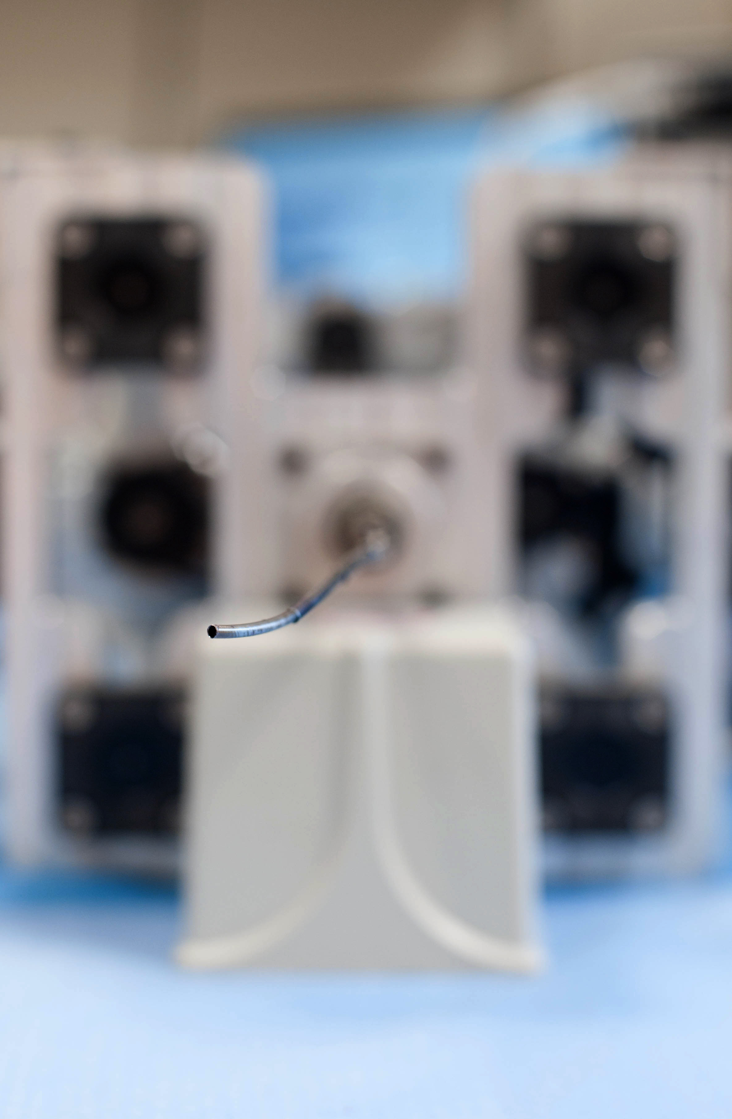
The business end of the robot, consisting of a series of concentric tubes, melts a simulated acoustic neuroma. The brain model, made through 3D printing, is based on MRI data from an real-life surgical case.
“That was the start for me,” says Codd, who is now completing his neurosurgical residency at Massachusetts General Hospital. “We got to batting ideas back and forth.” Along with engineer Tomer Anor, PhD, in Madsen’s lab, they formed a core team later joined by several postdoctoral fellows and graduate students.
It turns out just three to four small concentric tubes, one emerging from the other, create all the curviness you need to maneuver in areas just millimeters in size. “All our work happens between 2 and 3 mm,” says Codd. “The spaces we’re maneuvering in can be even smaller than in the heart.”
With seed money from the Innovation Acceleration Program at Boston Children’s Hospital, the Brain Science Foundation and the Boston Children’s/MIT collaborative grant program, they bought hardware and built a tabletop prototype. In current testing, a simple joystick is used to advance the tip in three dimensions, with motors used to rotate the tip and translate the user’s hand motions into tiny, intricate movements.
3D printing meets brain anatomy
Codd and Anor are aiming to refine this interface and program the robot to perform custom neurosurgeries, informed by patients’ actual MRI scans. Using molten plastic and 3D printing techniques, they can turn the MRI coordinates of prior surgical patients into physical models of the relevant brain areas. They’re using these to test the robot—to see whether it could have performed the tasks Codd accomplished through open brain surgery.
Anor built his lab’s first 3D printer from open-source instructions. In about two hours, it can make a small item like a plastic mold that, in turn, is used to create fake tumors for the robot to burn away. Anor hopes to use the printer to fabricate the parts for a bigger 3D printer that can build an entire model brain.But the ultimate goal, once the engineering kinks are worked out, is to move the robot to animal models. Codd thinks this can happen over the next year, starting with relatively simple maneuvers through the brain’s natural fluid-filled cavities (ventricles) and navigating very small spaces at the base of the brain.
“Why make a large hole like a barn door and have surgery take 10 hours when you can drill a little hole and thread in a snake-like robot and have the operation take one hour and cause minimal injury?” Codd says. “I have stacks of MRIs of children on whom I’ve operated. I go through them now and think, ‘maybe there is a way to do this better.’”